Views/Visit: 806
Case 20 : Film quiz : A 12 year old girl with incidental mass detected from chest radiograph.
Panruethai Trinavarat,M.D.
Assistant Professor.
Department of radiology, Faculty of medicine,
Chulalongkorn University
Case 20 :
Film quiz :
A 12 year old girl with incidental mass detected from chest radiograph.
Findings:
- A large lobulated mass (9.5x6 cm) at right lower hemithorax, silhouette with posterior two-thirds of right diaphragm, containing a dense oval calcification inferiorly.
- No pleural effusion.
- No adjacent or other infiltration.
- Normal cardiac size and normal pulmonary vasculature
- No evidence of hilar or mediastinal lymphadenopathy.
- Normal bony structure.
Opinion:
1) The mass could be originated from the lung, posterior mediastinum, and pleura.
2) Pleural mass is unlikely because it is almost always associated with pleural effusion.
3) For the lung mass with calcification, the differential diagnosis includes inflammatory pseudotumor, hamartoma, granuloma, pleuropulmonary blastoma, metastasis of some malignancies.
4) For the mediastinal mass with calcification, the differential diagnosis includes tumors of sympathetic ganglion origin (ganglioneuroma, ganglioneuroblastoma, and neuroblastoma).
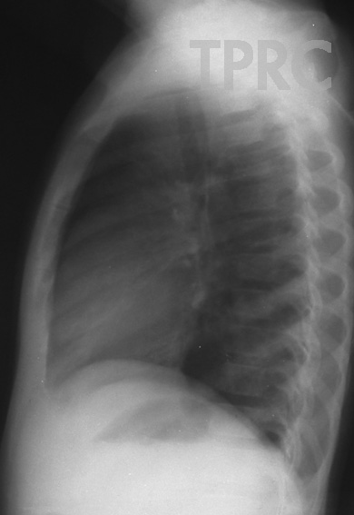
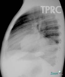
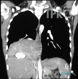
Figure 1.3:
Coronal plane reformation of arterial-phased CT chest at level of descending aorta
- Arterial phased CT chest is performed to detect origin of arterial supply to the mass. The study reveals right descending pulmonary artery and right inferior phrenic artery as two feeders. We may suggest the lesion starts from the lung, then increases size until attaching or invading mediastinum and it later gains the second artery supply from systemic circulation. On the contrary, mediastinal mass even very large size, will not gain blood supply from pulmonary circulation.
Opinion:
Final diagnosis from histology: Plasma cell granuloma
Inflammatory pulmonary pseudotumor, plasma cell granuloma, xanthogranuloma, and fibrous xanthoma are terms used to describe reactive myofibroblastic inflammatory process seen in children. It accounts for about half of benign lung tumors of childhood. The lesion is usually clinically silent.
Radiographs typically show a variably sized rounded or lobulated pulmonary parenchymal mass. Many of these lesions are more than 4 cm in diameter when detected. Calcification is common, often amorphous and scattered.
(Ref. Kird DR. Practical Pediatric Imaging Diagnosis Radiology of Infants and Children, 3rd ed. 1998; p 795)