
Pulmonary sequestration
Case 29 :
ผู้ป่วย เพศ หญิง อายุ 12 ปี จ.กรุงเทพฯ
admit ที่ รพ.นพรัตน์ราชธานี เมื่อ 7 กรกฎาคม 2547 ด้วยเรื่อง ไข้ ไอ เสมหะเขียว 1 wk
PE: BW 47 kg, p 100/min, RR 25/min, BT 36.2OC, decrease breath sound RLL
CXR: Imp: Lung abscess
Past Hx: ก.พ. 47 admit ด้วยเรื่อง Rt pleural effusion Rx ceftriaxone, Erythro 10days
มี.ค. 48 Dx pneumonia ไม่มี effusion
Hb10.5 Hct 34.6% WBC 22,330 N 87 L 7 M 6 Plt 473,000
CXR: air fluid level RLL
Sputum AFB mg x 3D, TT 12 mm
CT chest: pulmonary sequestration

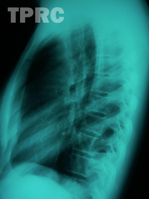
Chest : PA upright (b1)
- Cystic lesion with air-fluid level at medial part of right lower lung, neither silhouette with right heart border or the diaphragm. Lower extension of the opacity is to lung base; cannot tell whether there are more than one cysts or there is also solid component. Chest: Right lateral (b2)
- Posterior location of the lesion with air-fluid level; broad pleural-based lesion. DDX: Pulmonary cystic lesion: CCAM, pulmonary sequestration Pleural lesion : loculated pleural fluid, post tapping (so the lesion contains air)
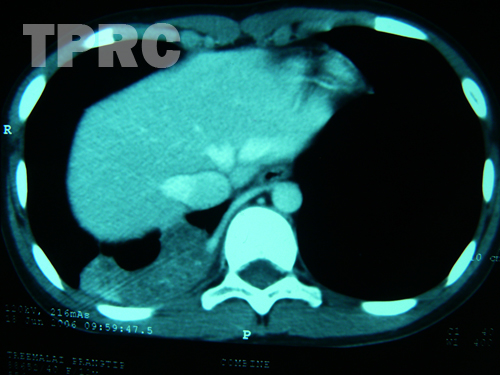
CTA of the thoracic aorta
(b3): Post IV contrast, axial plane at lung base, mediastinal window - A branch from right lateral wall of thoracoabdominal aorta feeds the pulmonary lesion in posterior segment of right lower lobe. The inferior solid-appearance portion of the pulmonary lesion with heterogeneous enhancement is noted.
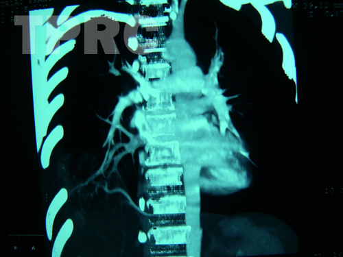
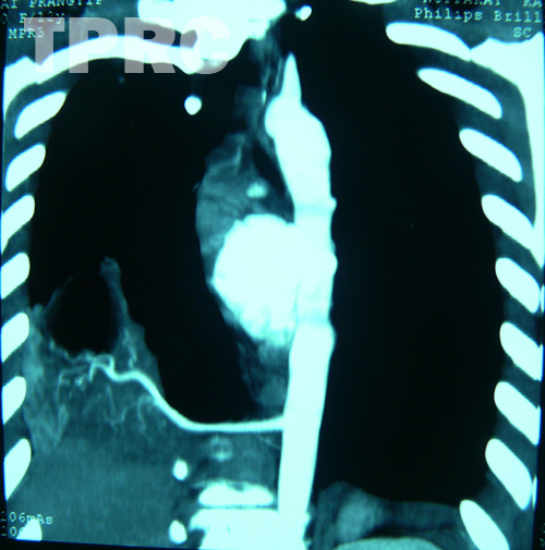
(b4 and b5): Reformatted MIP (maximal intensity projection) image in mildly-oblique coronal plane. - This view better demonstrates the arterial branch from thoracoabdominal aorta about level of diaphragm to supply complex solid-cystic lesion in right lower lobe, and venous drainage into right lower pulmonary vein.
DX: Intralobar pulmonary sequestration
สมาคมโรคระบบหายใจและเวชบำบัดวิกฤตในเด็กแห่งประเทศไทย
สำนักงาน: หน่วยโรคระบบหายใจเด็ก ชั้น 3 ห้อง 304 อาคารศูนย์แพทย์สิริกิต์
โรงพยาบาลรามาธิบดี พญาไท กรุงเทพมหานคร 10400
โทร. 0635894599
E-mail: thaipedlung.org@gmail.com