
CCAM
Case 28 :
ผู้ป่วย อายุ 2 เดือน refer จาก รพ.อ่างทองด้วยเรื่อง ไอ หายใจเร็ว
พบ pneumatocele, poor wt gain
ตรวจร่างกาย Lt 51, BW 2.3
Lung: rhonchi
CBC 6/6/50 Hct 32.4, Hb 10.9, WBC 10700 (N:7, LL 77, E7, M5, ATL 4) plt 548,000
HRCT = one large cyst and cluster ที่ multiple small cysts at Lt lower lung
UGIS: mod to severe GER, gastric wash for AFB XIII = negative
การวินิจฉัย LBW, lung cyst LLL, moderate to severe GER
การรักษาที่สำคัญ Ceftazidime + amilkin x 14 days (Rx sepsis)
Ranidine ,cisapride
การดำเนินโรค หลัง antibiotic x 14 d repeat H/C = NG
F/U CVT 2 wk


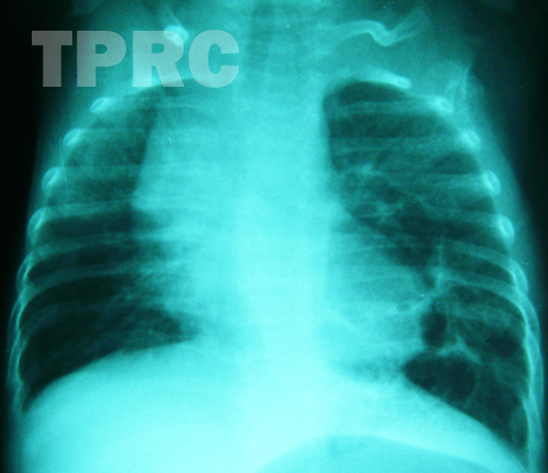
Chest : AP supine (a1)
- A group of cysts of variable sizes at medial side of left lower lobe. The largest cyst is above 2 cm size.
- Mildly increased volume of the left lung with flattening of left hemidiaphragm.
Intact diaphragm.
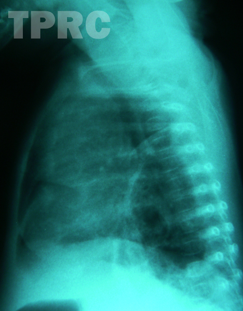
Chest : Left lateral (a2)
- Posterior location of the cysts with anterior displacement of pulmonary vessels in lower lung.
DDX: CCAM type 1, and pulmonary sequestration.
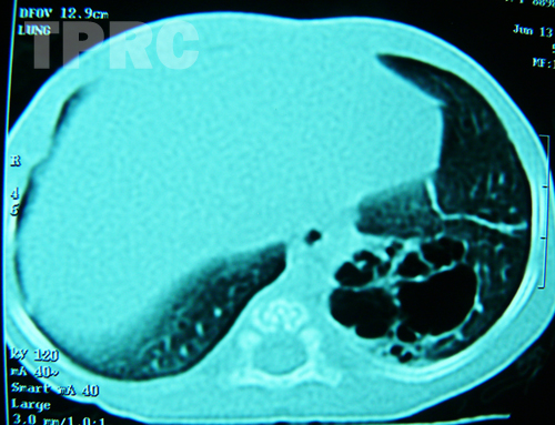
CT chest (a3): Axial plane of lower chest, using lung window.
- A group of small cysts in posterior basal segment of left lower lobe.
(Other images (not shown) using mediastinal window do not show systemic artery supply to this area of the lung.) DX: CCAM type 1
สมาคมโรคระบบหายใจและเวชบำบัดวิกฤตในเด็กแห่งประเทศไทย
สำนักงาน: หน่วยโรคระบบหายใจเด็ก ชั้น 3 ห้อง 304 อาคารศูนย์แพทย์สิริกิต์
โรงพยาบาลรามาธิบดี พญาไท กรุงเทพมหานคร 10400
โทร. 0635894599
E-mail: thaipedlung.org@gmail.com