
Panruethai Trinavarat,M.D.
Assistant Professor.
Department of radiology, Faculty of medicine,
Chulalongkorn University
Case 19 :
Film quiz:
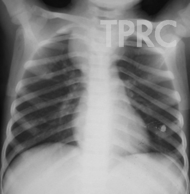
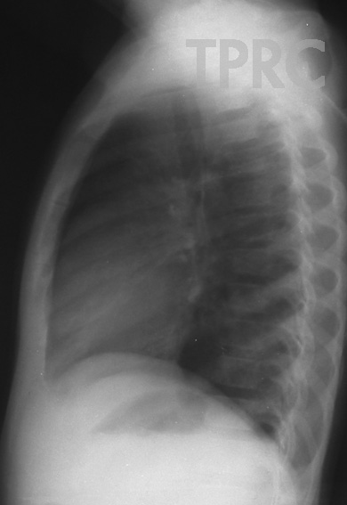
A 8 year-old boy with fever, weight loss, and anemia.
Findings:
- Superior mediastinal mass with bilateral convex bulging of the mediastinum, more on the right side. No calcification within the lesion.
- From lateral view, mildly increased opacity posterosuperiorly behind trachea.
- No tracheal shift in both PA and lateral views.
- Widening right and left 2nd-3rd intercostals spaces at posterior aspect, as compare to intercostals spaces at other levels.
- Normal heart, lungs, and pulmonary vasculature.
Opinion:
- Widening of intercostal space posteriorly in this case of mediastinal mass suggests the mass to be in posterior location, likely paraspinal mass with mass effect on the adjacent ribs.
- Differential diagnosis of posterior mediastinal mass in a child includes tumors of sympathetic ganglion origin (ganglioneuroma, ganglioneuroblastoma, neuroblastoma), nerve sheath tumor (neurofibroma, schwannoma), enteric or neurenteric cyst, lymphadenopathy.
- Further imaging investigation is MRI or CT.
MRI is preferred to CT in case of posterior mediastinal mass, because of better demonstration of intraspinal tumor extension which is common in neurogenic tumor.
Histologic diagnosis : Neuroblastoma
His bone survey shows multiple bone metastases.
Figure 1.1 and 1.2 : Chest PA(a) and left lateral (b) views


สมาคมโรคระบบหายใจและเวชบำบัดวิกฤตในเด็กแห่งประเทศไทย
สำนักงาน: หน่วยโรคระบบหายใจเด็ก ชั้น 3 ห้อง 304 อาคารศูนย์แพทย์สิริกิต์
โรงพยาบาลรามาธิบดี พญาไท กรุงเทพมหานคร 10400
โทร. 0635894599
E-mail: thaipedlung.org@gmail.com