
ผ.ศ.พญ.อาภัสสร วัฒนาศรมศิริ
Panruethai Trinavarat,M.D.
Assistant Professor.
Department of radiology, Faculty of medici
Case 18 :
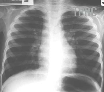
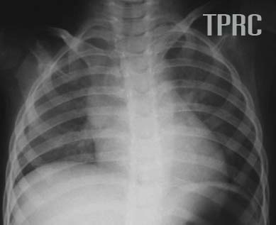
Figure 3:
expiratory chest (a) and inspiratory chest (b) and radiograph
Findings:
(a) Expiratory chest with posterior aspect of right 7th rib being above dome of right hemidiaphragm, narrowing intercostals spaces, buckling trachea, and sunken cardiac apex within the left hemidiaphragm. Much tracheal buckling that one may suspicious of left paratracheal mass. No definite pulmonary infiltration
(b) Good lung inflation with posterior aspect of right 9th rib being above dome of right hemidiaphragm, widening intercostal spaces, and straight trachea. Normal other findings but thickening of perihilar lung markings, probable from bronchitis, or asthma.


สมาคมโรคระบบหายใจและเวชบำบัดวิกฤตในเด็กแห่งประเทศไทย
สำนักงาน: หน่วยโรคระบบหายใจเด็ก ชั้น 3 ห้อง 304 อาคารศูนย์แพทย์สิริกิต์
โรงพยาบาลรามาธิบดี พญาไท กรุงเทพมหานคร 10400
โทร. 0635894599
E-mail: thaipedlung.org@gmail.com