
ผศ.พญ.อาภัสสร วัฒนาศรมศิริ
Panruethai Trinavarat,M.D. Assistant Professor.
Department of radiology, Faculty of medicine,
Case 25 :
ผู้ป่วยมีไข้สูง ไอ หายใจเหนื่อย
PE : subcostal retraction
Generalized exp. Wheezing, coarse crepitation
CBC : Hct 37% WBC 8,300 PMN 59 L 28
Mycoplasma IgM
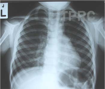
CHEST : PA, upright If the technician correctly put the marker (L) on patient’s left side, the patient should have situs inversus totalis. Cardiac size is normal. Mild perihilar and both lower lobe peribronchial opacities are seen. There is a small patchy opacity with sharp margin in left retrocardiac region, probable (sub)segmental atelectasis. If the patient has clinical suspicion of pneumonia, viral pneumonia is likely.

สมาคมโรคระบบหายใจและเวชบำบัดวิกฤตในเด็กแห่งประเทศไทย
สำนักงาน: หน่วยโรคระบบหายใจเด็ก ชั้น 3 ห้อง 304 อาคารศูนย์แพทย์สิริกิต์
โรงพยาบาลรามาธิบดี พญาไท กรุงเทพมหานคร 10400
โทร. 0635894599
E-mail: thaipedlung.org@gmail.com