
ผศ.พญ.อาภัสสร วัฒนาศรมศิริ
Panruethai Trinavarat,M.D.
Assistant Professor.
Department of radiology,
Case 22 :
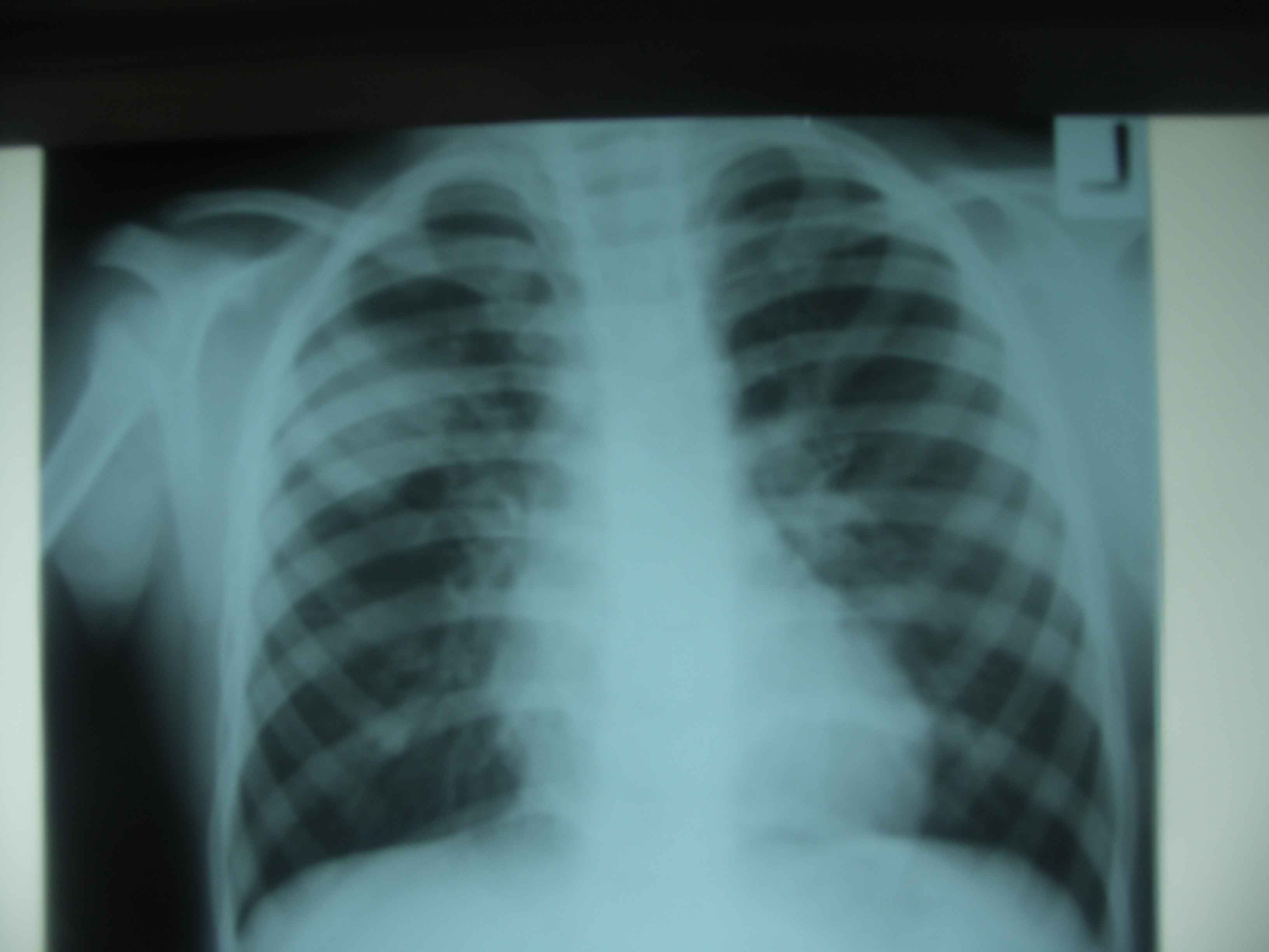
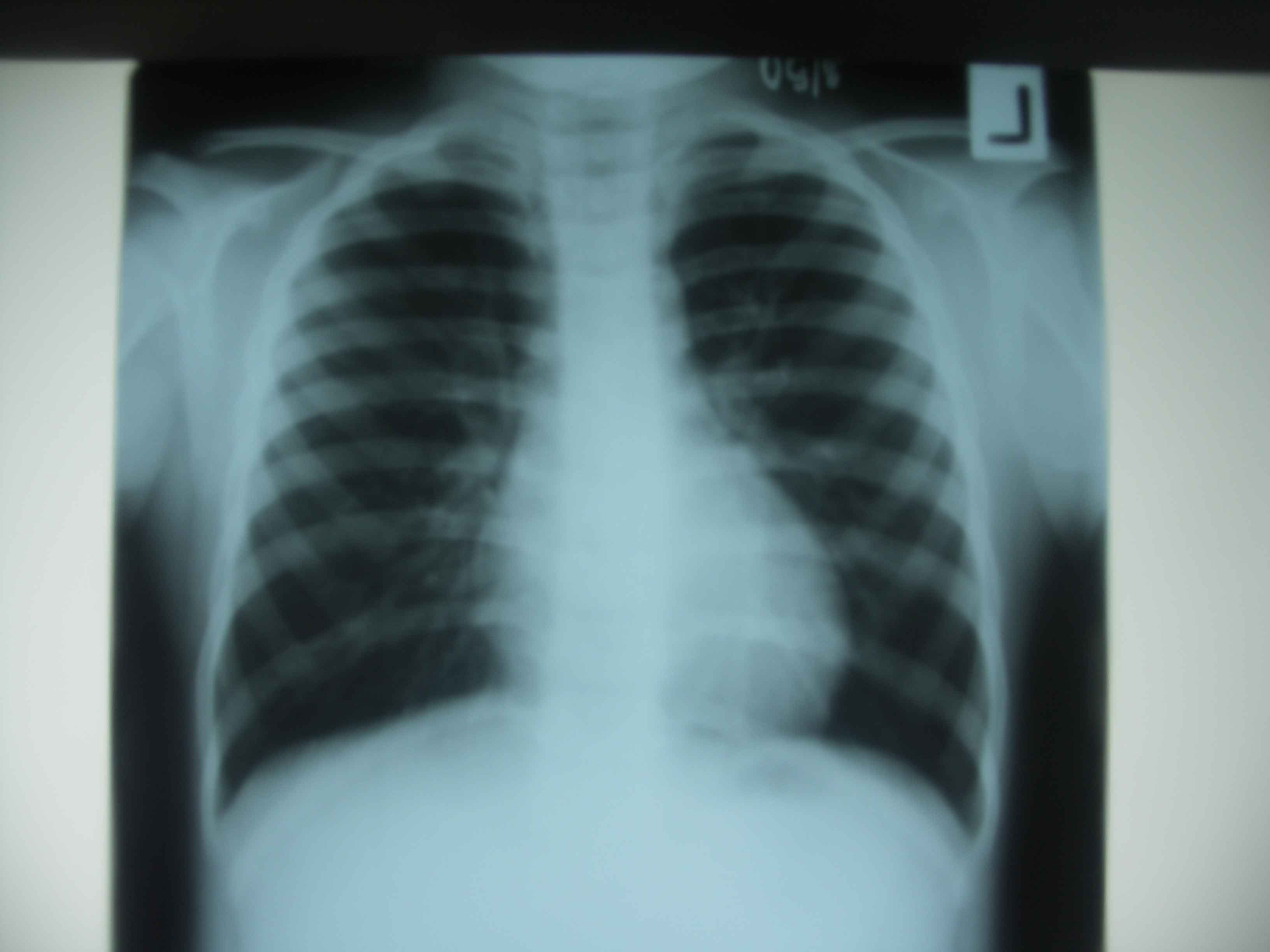
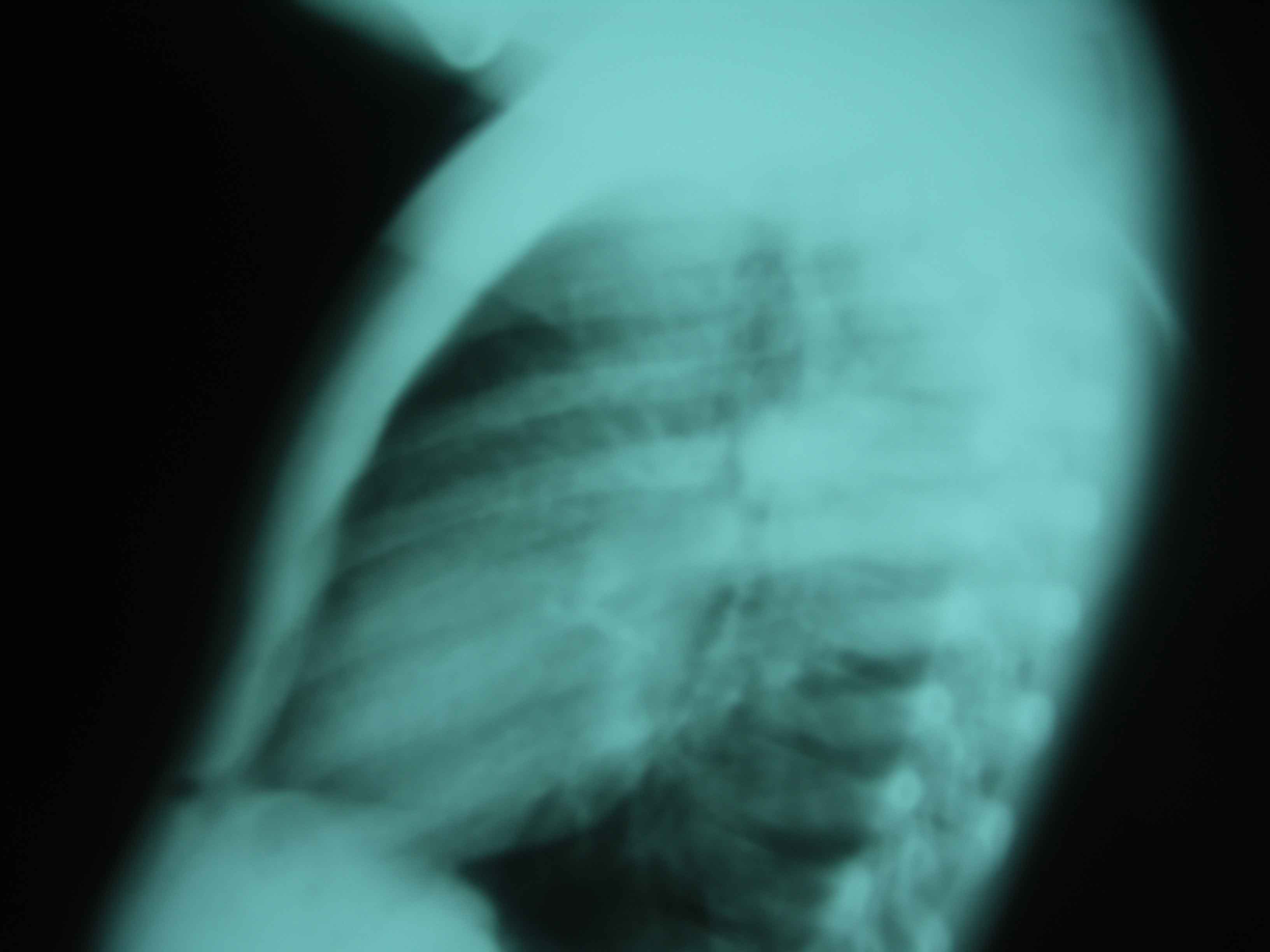
2- There is a mass-like lesion with mildly lobulated contour in right upper lung in PA view. In lateral view 2.1, it is suggested to be posteriorly located, possible in superior segment of right lower lobe. No pleural effusion or adenopathy is seen.
- With history of acute high fever, finding is likely from round pneumonia. But in other clinical settings, lung mass should be considered, such as metastasis, pleuropulmonary blastoma, hamartoma, intrapulmonary bronchogenic cyst.
- Because of its peripheral location in posterior aspect, ultrasound may have a role in differentiation between pneumonia and mass. Follow up chest radiograph will also help in confirming the diagnosis.
2.2 pictures are F/u CXR after treatment with ATB and supportive treatment


สมาคมโรคระบบหายใจและเวชบำบัดวิกฤตในเด็กแห่งประเทศไทย
สำนักงาน: หน่วยโรคระบบหายใจเด็ก ชั้น 3 ห้อง 304 อาคารศูนย์แพทย์สิริกิต์
โรงพยาบาลรามาธิบดี พญาไท กรุงเทพมหานคร 10400
โทร. 0635894599
E-mail: thaipedlung.org@gmail.com