
ผศ.พญ.อาภัสสร วัฒนาศรมศิริ
Panruethai Trinavarat,M.D. Assistant Professor.
Department of radiology, Faculty of medicine,
Case 24 :
ผู้ป่วยเด็กหญิง อายุ 11 ปี เคยหอบหลายครั้งตอนเด็ก ไม่หอบมาหลายปี ครั้งนี้ไอมีเสมหะ น้ำมูก และเหนื่อยมา 2 วัน ก่อนมาโรงพยาบาล ไม่ได้พาไปพบแพทย์ วันที่พามาสังเกตว่าไอเหนื่อยมาก ไม่มีประวัติ choking
PE : marked congest nasal mucosa with mucoid discharge SpO2 room air 93%, subcostal retraction Breath sound of Lt. lung
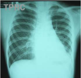
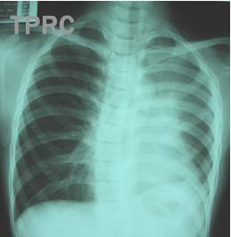
CHEST: PA upright In both films, there is decreased left lung volume with mediastinal shift to the left and elevation of left hemidiaphragm, more in image B. In A, the left retrocardaic area is opaque and border of the left hemidiaphragm is poorly seen; possible from consolidation or atelectasis. With evidence of volume loss, LLL atelectasis is favorable, even the typical sharp lateral demarcation line is not seen. In B, the upper part of left cardiac border and pulmonary trunk are obliterated with opacity of left upper lung, and evidence of volume loss of the left lung; indicating LUL atelectasis.


สมาคมโรคระบบหายใจและเวชบำบัดวิกฤตในเด็กแห่งประเทศไทย
สำนักงาน: หน่วยโรคระบบหายใจเด็ก ชั้น 3 ห้อง 304 อาคารศูนย์แพทย์สิริกิต์
โรงพยาบาลรามาธิบดี พญาไท กรุงเทพมหานคร 10400
โทร. 0635894599
E-mail: thaipedlung.org@gmail.com