
Panruethai Trinavarat,M.D.
Assistant Professor.
Department of radiology, Faculty of medicine,
Chulalongkorn University
Case 1 :
Question :
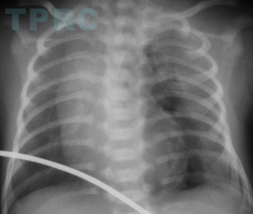
A 5 day old boy with respiratory distress. What is the diagnosis?
Answer :
The medial pneumothorax, left
Pneumothorax on chest radiograph is usually seen as air density lateral to or above the displaced lung. However, in neonates and infants who are maintained in supine position, air in pleural space is preferably anteriorly and medially located between the medial surface of the lung and the anterior mediastinum.

Chest radiograph in supine position will show a dark band of air between the lung and mediastinum, the “sharp mediastinum” sign in medial pneumothorax. If there is large amount of the pneumothorax, contralateral mediastinal shift will be seen.
Intercostal drainage for this type of pneumothorax should be performed with directing the tip of pleural tube anteriorly.
สมาคมโรคระบบหายใจและเวชบำบัดวิกฤตในเด็กแห่งประเทศไทย
สำนักงาน: หน่วยโรคระบบหายใจเด็ก ชั้น 3 ห้อง 304 อาคารศูนย์แพทย์สิริกิต์
โรงพยาบาลรามาธิบดี พญาไท กรุงเทพมหานคร 10400
โทร. 0635894599
E-mail: thaipedlung.org@gmail.com