
Loculated effusion
Case 36 :
เด็กหญิงอาหรับ อายุ 3 ปี ภูมิลำเนา ประเทศ United Arab Emirates(UAE)


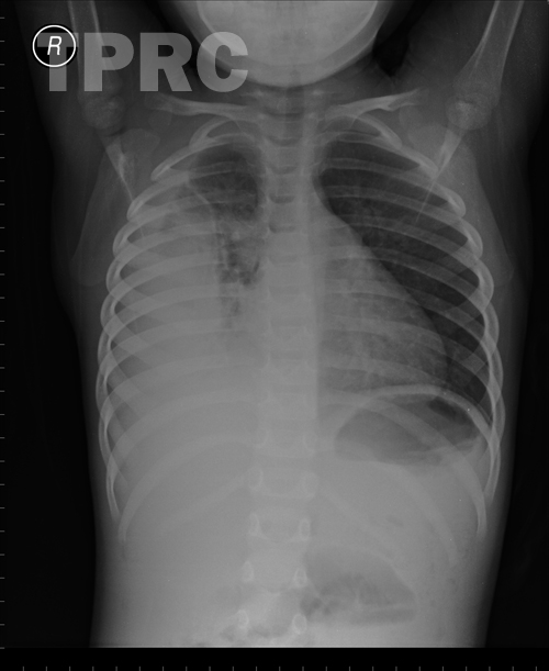
Chest: AP supine
- Homogeneously-dense patchy opacity is noted in right upper lobe with sharply defined linear inferior border with mildly downward position of minor fissure. Impression: Consolidation (alveolar infiltration) in anterior segment of right upper lobe; possible pneumonia (bacterial pattern), pneumonitis (according to history), or hemorrhage.
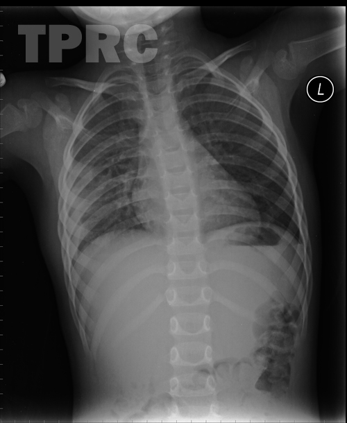
Chest: PA, upright (i1); post treatment - Much decreased size of the opacity in the right lung, with remained loculated pleural effusion (or pleural thickening) at lateral and posterior aspects of right lower hemithorax.
สมาคมโรคระบบหายใจและเวชบำบัดวิกฤตในเด็กแห่งประเทศไทย
สำนักงาน: หน่วยโรคระบบหายใจเด็ก ชั้น 3 ห้อง 304 อาคารศูนย์แพทย์สิริกิต์
โรงพยาบาลรามาธิบดี พญาไท กรุงเทพมหานคร 10400
โทร. 0635894599
E-mail: thaipedlung.org@gmail.com