
Bronchogenic cyst
Case 31 :
เด็กหญิงไทยใหญ่อายุ 8 เดือน ภูมิลำเนา อ.แม่ลาน้อย จ.เชียงใหม่ ไข้ ไอหายใจเหนื่อย 3 วันก่อนมารพ


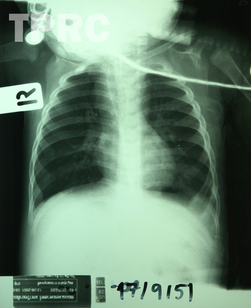
Chest: AP supine (d1)
- Bilateral pulmonary hyperinflation.
- Minimal infiltration in lateral aspect of left lung base.
- Mildly convex bulging both right and left sides of superior mediastinum, more on the right, with prominent shift of NG tube to the right IMPRESSION ; Superior mediastinal mass, suggested to be posterior compartment.
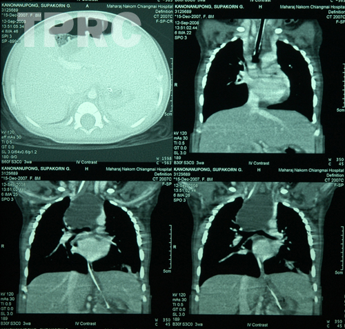
CT chest: post IV contrast
(d2) A series of 3 reformatted coronal plane at level of trachea and behind.
- A large oval shaped non-enhanced hypodense mass with smooth and sharp margins in superior mediastinum behind the trachea.
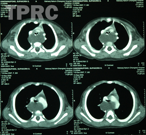
(d3) A series of 4 axial plane at level of aortic arch and above.
- Wide separation of trachea and esophagus by the mediastinal hypodense mass.
- Esophagus with indwelling NG is noted at right paraspinal area.
IMPRESSION; Mediastinal mass, suggestive to be uniloculated cystic lesion, locates between middle and posterior mediastinum.
DDX: Bronchogenic cyst VS enteric cyst.
Cystic hygroma may also be in DDX but more commonly cystic hygroma is multiloculated.
สมาคมโรคระบบหายใจและเวชบำบัดวิกฤตในเด็กแห่งประเทศไทย
สำนักงาน: หน่วยโรคระบบหายใจเด็ก ชั้น 3 ห้อง 304 อาคารศูนย์แพทย์สิริกิต์
โรงพยาบาลรามาธิบดี พญาไท กรุงเทพมหานคร 10400
โทร. 0635894599
E-mail: thaipedlung.org@gmail.com