
Lung abscess
Case 32 :
เด็กหญิงไทยใหญ่อายุ 8 เดือน ภูมิลำเนา อ.แม่ลาน้อย จ.เชียงใหม่ ไข้ ไอหายใจเหนื่อย 3 วันก่อนมารพ... 3 เดือนก่อนมาโรงพยาบาล ผู้ป่วยเริ่มมีอาการไข้เป็น ๆ หาย ๆ


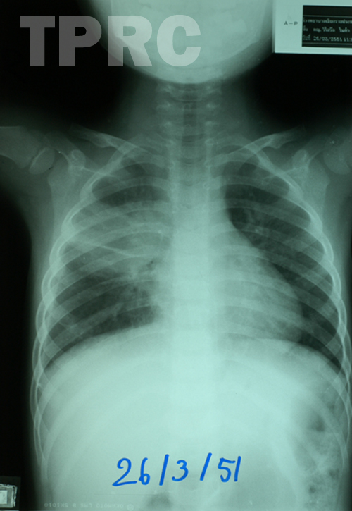
Chest: AP supine (e1) : 26/3/51
- Mass like consolidation in right upper and middle chest, crossing minor fissure which is mildly thickened.
Impression ; Pneumonia of alveolar pattern in superior segment of right lower lobe.
Minimal right pleural effusion.
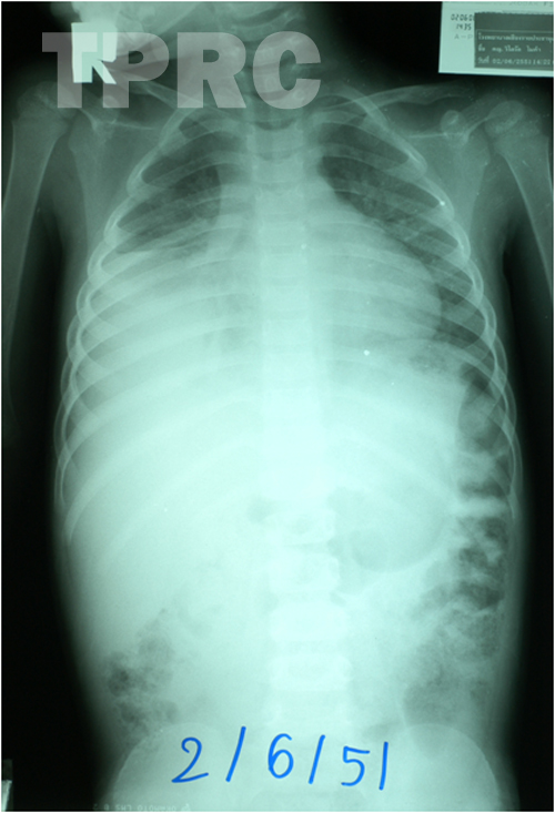
Chest: AP supine (e2) : 02/6/51
- Large consolidated area in lower half of right lung with remained small aerated lung close to right heart borer.
Impression ; Pneumonia of alveolar pattern in right lower lobe and right middle lobe.
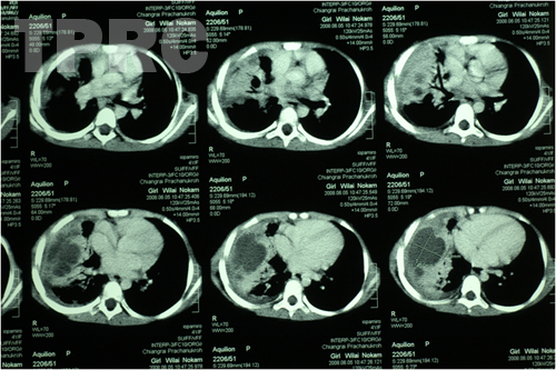
CT chest: a series of 6 axial images in lower half of the chest (e3) : 05/6/51
- Multiple discrete and coalesce hypodense lesions, some with enhanced walls, within the areas of pulmonary consolidation in right middle and right lower lobes.
- Minimal free right pleural effusion seen in dependent portion of the chest in lower images.
Impression : Multiple pulmonary abscesses in lobar consolidation of right middle and lower lobes.
สมาคมโรคระบบหายใจและเวชบำบัดวิกฤตในเด็กแห่งประเทศไทย
สำนักงาน: หน่วยโรคระบบหายใจเด็ก ชั้น 3 ห้อง 304 อาคารศูนย์แพทย์สิริกิต์
โรงพยาบาลรามาธิบดี พญาไท กรุงเทพมหานคร 10400
โทร. 0635894599
E-mail: thaipedlung.org@gmail.com