
superior segment pneumonia-atelectasis
Case 38 :
ผู้ป่วยอายุ 9 ปี แข็งแรงดี ไข้สูงมา 3 วัน ไปโรงพยาบาลได้ Amoxicillin + Roxithromycin ไม่ดีขึ้น


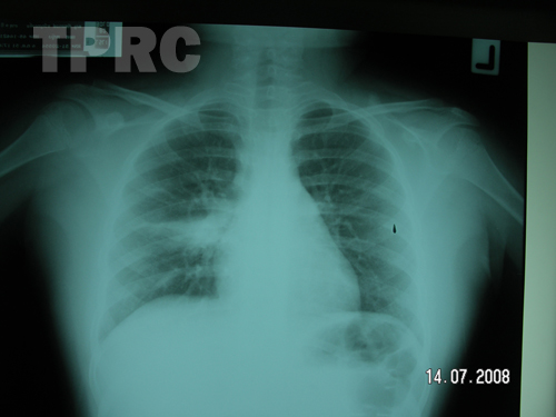
Chest: PA upright (k1)
- A triangular shape opacity is noted at middle part of right lung with its base towards the mediastinum but not silhouette with right heart border. Its upper margin is sharply defined in linear outline. Impression; Suspecious of atelectasis of right middle lobe (from its shape and sharp linear upper margin); however, unusual for non-sillhouette with right border, probable a part of medial segment is spared from atelectasis.
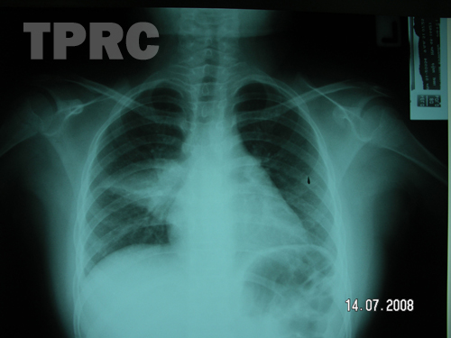
Chest: PA upright (k2)
- Follow up film shows larger size of the triangular shape lesion with now sharp inferior margin. The minor fissure appears to be seen as at thick linear opacity superimposed on the lesion. Right heart border is still well seen.
Impression; Pulmonary atelectasis is possible in superior segment of right lower lobe, not in middle lobe as previously considered.
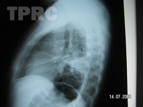
Chest: Right lateral (k3)
- Lateral view confirms triangular-shaped opacity in superior segment of right lower lobe, with sharp linear outline of major fissure which is downwards in position. Inferior margin of the atelectasis also has sharp linear margin.
Impression; Atelectasis of superior segment of right lower lobe.
สมาคมโรคระบบหายใจและเวชบำบัดวิกฤตในเด็กแห่งประเทศไทย
สำนักงาน: หน่วยโรคระบบหายใจเด็ก ชั้น 3 ห้อง 304 อาคารศูนย์แพทย์สิริกิต์
โรงพยาบาลรามาธิบดี พญาไท กรุงเทพมหานคร 10400
โทร. 0635894599
E-mail: thaipedlung.org@gmail.com