
LUL agenesis
Case 40 :
ผู้ป่วยเด็กหญิงไทย อายุ 5 เดือน มีอาการไข้ ไอ หายใจหอบไปรักษาที่ รพ. รัฐ วินิจฉัยปอดบวม ให้ยาปฏิชีวนะ นอน รพ. 5 วัน
3 วันก่อน มีอาการไข้ ไอ น้ำมูกใส หายใจครืดคราด หอบ
ตรวจร่างกาย –RR 40/min, T 37.5 oC No cyanosis, Tachypnea
HEENT- pharynx mild injected. Nasal discharge clear
Heart normal s1s2 no murmur
Lung- asymmetrical chest wall, transmitted sound, no wheezing, asymmetrical breathsound
CBC- Hb 10.3 g/dl, Hct 32.1%, Wbc 10,500/cumm. PMN 14% Lym 80 Mo 6 plt
Adequate
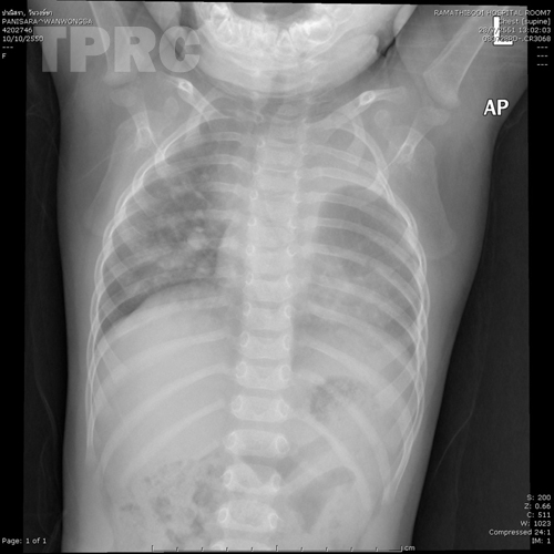
Chest: AP supine (m1)
- Small left lung volume.
- Small and mildly deformed left 3rd rib.
- Opacity in left side of superior mediastinum continued to upper one-fourth portion of left hemithorax with sharply-outlined inferior concave margin.
- Tracheal shift to the right, uncertained whether caused by left sided superior mediastinal lesion or suboptimal inspiration.
- Partially obscured border of left hemidiaphragm, and left cardiac border.
- Increased flow to right lung, and relatively decreased flow to left lung.
- Normal cardiac size.
Impression: Small left lung volume, probable from congenital or atelectasis.
Left sided superior mediastinal mass, uncertain whether it is true mass or thymus; further investigation with US or CT is recommended.
Asymmetrical pulmonary flow to right and left lungs.
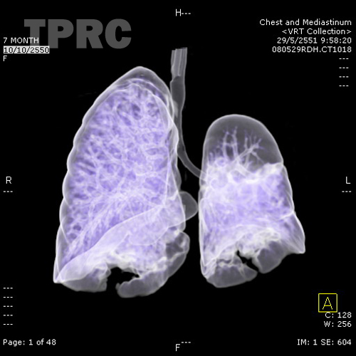
CT chest: Volume rendering technique of the lung and tracheobronchial tree in frontal projection (m2)
- Small size of the left lung, and no atelectasis. Other images (not shown) revealed agenesis of left upper lobe.
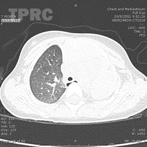
CT chest: Axial plane at level of upper chest (m3)
- Aerated lung only in right upper lobe, but not in left upper chest. Mediastinal window (not shown) shows normal thymic tissue in left upper chest.
สมาคมโรคระบบหายใจและเวชบำบัดวิกฤตในเด็กแห่งประเทศไทย
สำนักงาน: หน่วยโรคระบบหายใจเด็ก ชั้น 3 ห้อง 304 อาคารศูนย์แพทย์สิริกิต์
โรงพยาบาลรามาธิบดี พญาไท กรุงเทพมหานคร 10400
โทร. 0635894599
E-mail: thaipedlung.org@gmail.com